

Introduction: Prostate cancer (PCa) stands as the most prevalent non-cutaneous malignancy among males, with significant incident cases and mortality rates in the United States alone. Advancements in treatment, particularly in radiation therapy (RT), have paved the way for more precise and effective modalities. Among these, intensity-modulated radiotherapy (IMRT) and stereotactic body radiotherapy (SBRT) have emerged as leading choices for localized PCa. However, the efficacy of these treatments hinges on addressing a critical concern—geometric uncertainty.
Understanding Geometric Uncertainty: Geometric uncertainty in RT planning and delivery encompasses errors in target delineation, patient setup, interfraction, and intrafraction motion. The distinction between systematic and random errors plays a crucial role. Systematic errors occur during planning, while random errors vary with each fraction. Target delineation errors, arising from imaging modality limitations and observer variability, can significantly impact treatment outcomes.
Patient setup errors, another source of uncertainty, highlight the importance of immobilization techniques. Techniques such as pelvic and leg immobilization have been explored to enhance the reproducibility of treatment setup. Additionally, target position variation, both in terms of day-to-day interfraction motion and intrafraction movement during treatment, adds complexity. Studies have demonstrated the correlation between rectal filling and prostate position variability, emphasizing the need for comprehensive strategies to manage these uncertainties.
The Role of Image-Guided Radiation Therapy (IGRT): To mitigate the risks associated with geometric uncertainty, Image-Guided Radiation Therapy (IGRT) emerges as a vital tool. IGRT allows for daily adjustments to patient setup and real-time correction of radiation beams during delivery. This not only improves treatment accuracy but also enables a reduction in planning margins, thereby minimizing normal tissue toxicity.
Techniques Addressing Geometric Uncertainty: Various IGRT modalities are employed to address geometric uncertainty. Technologies such as cone-beam computed tomography (CBCT), implanted fiducial markers (FMs), ultrasound, and magnetic resonance imaging (MRI) play pivotal roles. CBCT offers excellent spatial resolution for daily positioning, while FMs, like gold seeds or coils, provide a low-cost solution for daily in-room imaging. MRI-guided RT stands out for its ability to track the target during treatment.
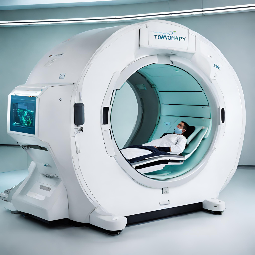
- Tomotherapy based IGRT
In the dynamic landscape of medical technology, advancements in radiation therapy have been crucial in enhancing the precision and effectiveness of cancer treatments. One such groundbreaking technique is Tomotherapy, a cutting-edge form of intensity-modulated rotational radiation therapy (RT) using a photon fan beam. In this blog post, we’ll explore the intricacies of Tomotherapy, with a particular focus on its helical variant, shedding light on its benefits and the transformative impact it has on cancer treatment.
Understanding Tomotherapy:
Tomotherapy represents a paradigm shift in radiation therapy, offering a sophisticated approach to treating cancer with enhanced precision. Unlike conventional radiation therapy, Tomotherapy utilizes a fan beam of photons to deliver intensity-modulated radiation. This allows for a highly tailored dose distribution, optimizing the treatment’s efficacy while minimizing damage to surrounding healthy tissues.
Helical Tomotherapy: A Continuous Revolution:
At the forefront of Tomotherapy innovation is the helical variant, a technique characterized by the continuous motion of the treatment couch and gantry along a helical trajectory. This dynamic approach offers several advantages, including improved coverage and integrated image guidance for precise patient setup verification.
- Tailored Dose Distributions: The helical motion of the treatment apparatus enables the delivery of highly customized dose distributions. This tailoring ensures that the radiation is concentrated on the cancerous tissue while sparing healthy surrounding structures.
- Continuous Motion for Enhanced Precision: Unlike traditional radiation therapy, which involves static beams, helical tomotherapy’s continuous motion allows for a more precise and uniform delivery of radiation. This feature is particularly beneficial when treating tumors with complex shapes or those situated near critical structures.
- Image Guidance for Setup Verification: Helical tomotherapy integrates image guidance, providing real-time imaging during treatment. This allows clinicians to verify and adjust the patient’s setup, ensuring the accuracy of the radiation delivery.
Adaptive Planning: A Dynamic Approach to Treatment:
One of the distinctive features of helical tomotherapy is its incorporation of adaptive planning software. This software monitors changes in both external and internal anatomy throughout the course of treatment. If significant alterations are detected, the adaptive planning system can modify the radiation treatment plans in real-time. This capability enhances the treatment’s responsiveness to the dynamic nature of cancer and contributes to improved outcomes.
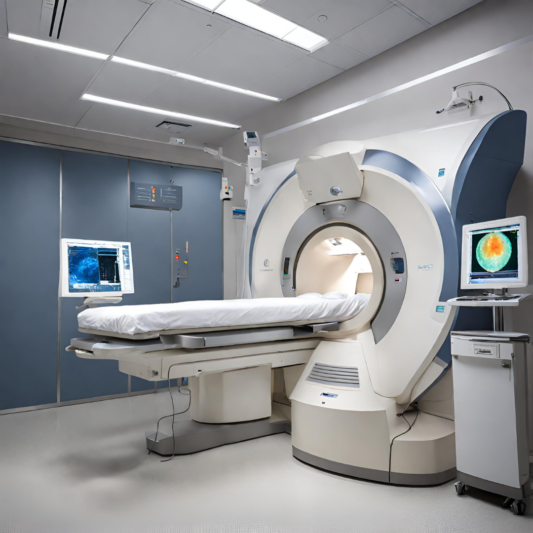
- Linac Based IGRT
Linear accelerator (linac) based Image-Guided Radiation Therapy (IGRT) is a widely used technique in modern radiation therapy. It combines the precision of image guidance with the versatility of a linear accelerator to deliver accurate and targeted radiation treatment.
Linac based IGRT utilizes onboard imaging systems, such as cone-beam computed tomography (CBCT), to acquire high-resolution images of the patient’s anatomy before each treatment session. These images are then compared to the reference images taken during the planning phase to ensure precise patient setup and target localization.
By incorporating IGRT into linac-based radiation therapy, clinicians can account for daily anatomical variations and make necessary adjustments to ensure accurate radiation delivery. This approach minimizes uncertainties caused by patient setup errors, organ motion, and changes in tumor size and shape throughout the treatment course.
Linac based IGRT offers numerous benefits, including improved treatment accuracy, reduced radiation toxicity to healthy tissues, and increased tumor control rates. With the ability to adapt treatment plans based on daily imaging, clinicians can confidently deliver precise radiation therapy to prostate cancer patients.
However, challenges exist, including motion artifacts, distortion, and limitations for patients with metallic implants.
Clinical Outcomes: IGRT’s Impact on Treatment Efficacy and Toxicity
The implementation of IGRT has significantly improved treatment accuracy and margin reduction in prostate cancer radiation therapy. While level 1 evidence is still evolving, historical data and recent studies provide compelling insights.
Studies comparing IGRT to non-IGRT cohorts demonstrate improved post-treatment quality of life, reduced acute treatment-related toxicity, and significant reductions in late urinary toxicity. The AIM trial, assessing the impact of margin reduction, reports decreased acute reduction in patient-reported quality of life outcomes with reduced PTV margins using IGRT.
Challenges and Considerations: While IGRT techniques offer unprecedented precision, challenges persist. Issues like marker migration and invasive procedures for marker placement must be considered. Furthermore, the choice of imaging modality, such as CT or MRI, introduces its own set of challenges related to accuracy and registration.
Conclusion: In navigating the complex landscape of prostate cancer radiation therapy, addressing geometric uncertainty through IGRT is paramount. Understanding the sources of uncertainty and leveraging advanced IGRT techniques empower clinicians to optimize treatment outcomes while minimizing the impact on surrounding healthy tissues. As technology continues to evolve, the role of IGRT in shaping the future of prostate cancer care remains pivotal.